Diagram Of Shoulder Muscles And Tendons / The rotator cuff is a group of four muscles and tendons that surround the glenohumeral joint.
Pablo-
0
Diagram Of Shoulder Muscles And Tendons / The rotator cuff is a group of four muscles and tendons that surround the glenohumeral joint.. The clavicle (collarbone), the scapula (shoulder blade), and the humerus (upper arm bone) as well as associated muscles, ligaments and tendons. Muscles of the shoulder work in team to produce highly coordinated motion. Learn vocabulary, terms and more with flashcards, games and other study tools. The core muscles are those in the abdomen, back, and pelvis, and they also stabilize the body and assist in tasks, such as lifting weights. Muscles move the bones by pulling on the tendons.
Muscle diagram leg 12 photos of the muscle diagram leg front leg muscle diagram, leg muscle diagram wikipedia, muscle anatomy of leg and foot, muscle diagram of upper leg, muscular diagram of leg. The rotator cuff tendons are a group of four tendons that connect the deepest layer of muscles to the humerus. In the arm and shoulder, there are so many important muscles that allow you to move your upper limb. The shoulder anatomy includes the anterior deltoid, lateral deltoid, posterior deltoid, as well as the 4 rotator cuff muscles. The capsule is strengthened by the tendons and ligaments surrounding and blending with it.
Anterior view of the chest and shoulder | Shoulder muscle ... from i.pinimg.com These muscles form the outer shape of the shoulder and underarm. The shoulder has about eight muscles that attach to the scapula, humerus, and clavicle. Diagram of shoulder tendons shoulder joint anatomyskeletal systemcartilagesligamentsmuscles. Muscles of the shoulder work in team to produce highly coordinated motion. Muscle tendons stretch over joints and contribute to joint stability. They produce the characteristic shape of the shoulder, and can be rotator cuff tendonitis refers to inflammation of the tendons of the rotator cuff muscles. Related posts of diagram of shoulder muscles and tendons muscle anatomy of chest. This usually occurs secondary to repetitive use of the shoulder.
Originates from the ulna, splitting into four tendons at the wrist which travel through the carpal tunnel and attach distally to the fingers.
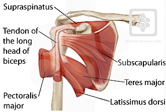
Originates from the ulna, splitting into four tendons at the wrist which travel through the carpal tunnel and attach distally to the fingers. Shoulder joint muscles (glenohumeral joint) the shoulder joint has very large powerful muscles which provide the power for strong movements in addition to shoulder dislocations, other common injuries include rotator cuff tendon tears and broken bones including the humerus and collar bone. Muscle tendons stretch over joints and contribute to joint stability. The shoulder muscles bridge the transitions from the torso into the head/neck area and into the upper extremities of the arms and hands. The shoulder is one of the largest and most complex joints in the body. Muscle tendons in the knee joint and the shoulder joint are crucial in stabilization. This usually occurs secondary to repetitive use of the shoulder. Starting point the muscles are the supraspinatus this is a flat triangular muscle that fills the entire infraspinatus fossa. Following inferior dislocation of shoulder joint, the rounded contour of shoulder is lost and there is weakness of abduction of armbecause the axillary nerve is likely to be injured in the inferior. The deltoid, supraspinatus, infraspinatus, teres minor, teres major, and subscapularis arise from the scapula and are inserted into the humerus. The human shoulder is made up of three bones: Muscles move the bones by pulling on the tendons. These muscles form the outer shape of the shoulder and underarm.
The muscles of the shoulder are associated with movements of the upper limb. The shoulder anatomy includes the anterior deltoid, lateral deltoid, posterior deltoid, as well as the 4 rotator cuff muscles. Related posts of shoulder muscles and tendons diagram. This usually occurs secondary to repetitive use of the shoulder. The shoulder muscles include skeletal muscles that are attached to the head of the humerus which performs various direct and indirect functions of the shoulder joints.
Shoulder / ПЛЕЧИ: Rotator Cuff Mechanics from www.shoulderdoc.co.uk Originates from the ulna, splitting into four tendons at the wrist which travel through the carpal tunnel and attach distally to the fingers. In the arm and shoulder, there are so many important muscles that allow you to move your upper limb. The rotator cuff tendons are a group of four tendons that connect the deepest layer of muscles to the humerus. Shoulder flexion is movement of the shoulder in a forward motion. The muscles in the shoulder aid in a wide range of movement and help protect and maintain the main shoulder joint, known as the. Major muscles the muscles that are responsible for movement in the shoulder attach to the scapula, humerus, and clavicle. Learn vocabulary, terms and more with flashcards, games and other study tools. Webmd's shoulder anatomy page provides an image of the parts of the shoulder and describes its function, shoulder problems, and more.
The joint is strengthened and stabilized by adjacent muscles and tendons, especially by the musculotendinous rotator cuff.
Each of these muscles is a discrete organ constructed of skeletal muscle tissue, blood vessels, tendons, and nerves. Related posts of diagram of shoulder muscles and tendons muscle anatomy of chest. Ready to test your knowledge on those muscles? The rotator cuff tendons are a group of four tendons that connect the deepest layer of muscles to the humerus. However, their origin is found in the osseous structures and they are not to be included with the rotator cuff muscles. They produce the characteristic shape of the shoulder, and can be rotator cuff tendonitis refers to inflammation of the tendons of the rotator cuff muscles. An example of shoulder flexion can be seen when reaching forward to grasp an object. Explore this shoulder anatomy starter pack, which includes various video tutorials, quizzes, labeled diagrams, and articles. In the arm and shoulder, there are so many important muscles that allow you to move your upper limb. Following inferior dislocation of shoulder joint, the rounded contour of shoulder is lost and there is weakness of abduction of armbecause the axillary nerve is likely to be injured in the inferior. Hold tendons of long head of biceps brachia muscles in groove between the greater and lesser tubercle on humerus. Recurring dislocations, which may be partial or complete, cause pain and unsteadiness when you raise your arm or move it away from your body. Muscle tendons in the knee joint and the shoulder joint are crucial in stabilization.
The shoulder is one of the largest and most complex joints in the body. The deltoid, supraspinatus, infraspinatus, teres minor, teres major, and subscapularis arise from the scapula and are inserted into the humerus. This usually occurs secondary to repetitive use of the shoulder. The capsule is strengthened by the tendons and ligaments surrounding and blending with it. Related posts of diagram of shoulder muscles and tendons muscle anatomy of chest.
Pin on OT from i.pinimg.com Muscle tendons in the knee joint and the shoulder joint are crucial in stabilization. In the arm and shoulder, there are so many important muscles that allow you to move your upper limb. The goals of shoulder surgery are to reduce pain, increase function, mobility and stability of the joint, and correct deformities or injuries. The shoulder muscles include skeletal muscles that are attached to the head of the humerus which performs various direct and indirect functions of the shoulder joints. The shoulder has about eight muscles that attach to the scapula, humerus, and clavicle. Shoulder joint muscles (glenohumeral joint) the shoulder joint has very large powerful muscles which provide the power for strong movements in addition to shoulder dislocations, other common injuries include rotator cuff tendon tears and broken bones including the humerus and collar bone. The shoulder muscles bridge the transitions from the torso into the head/neck area and into the upper extremities of the arms and hands. The capsule is strengthened by the tendons and ligaments surrounding and blending with it.
Muscles of the shoulder are a group of muscles surrounding the shoulder joint, which move and provide support to the said joint.
Specifically, the four rotator cuff muscles include the following For that reason, and because of the dexterity of the shoulder joint itself, the musculature of the shoulder is complex, ranging from massive prime mover muscles to. The shoulder is not a single joint, but a complex arrangement of bones, ligaments, muscles, and tendons that is better called the shoulder girdle. Muscles and tendons of the forearm and hand: The muscles of the shoulder are associated with movements of the upper limb. The rotator cuff tendons are a group of four tendons that connect the deepest layer of muscles to the humerus. This usually occurs secondary to repetitive use of the shoulder. An example of shoulder flexion can be seen when reaching forward to grasp an object. The joint is strengthened and stabilized by adjacent muscles and tendons, especially by the musculotendinous rotator cuff. Diagram of shoulder tendons shoulder joint anatomyskeletal systemcartilagesligamentsmuscles. Start studying shoulder ligaments and tendons. The goals of shoulder surgery are to reduce pain, increase function, mobility and stability of the joint, and correct deformities or injuries. The core muscles are those in the abdomen, back, and pelvis, and they also stabilize the body and assist in tasks, such as lifting weights.