Bone Cross Section Under Microscope / Cross section of ground compact bone.
Pablo-
0
Bone Cross Section Under Microscope / Cross section of ground compact bone.. To start, select the structure on the model. Bone cross section illustrations & vectors. Bones are rigid organs that support and protect various organs of the body, produce red and white blood cells and store minerals. Bone cross section — stock image & photo. The circular patterns are the concentric lamellae of the haversian canal in the center.
Millimeter circumferential area in which the bone figure 4. Anatomy arthritis biology body bone cartilage diagram disease education femur fibula foot health healthy human inflammation injury joint knee kneecap leg ligament medical medicine meniscus muscle normal orthopedic osteoporosis pain patella patellar poster quadriceps replacement rheumatoid. The jeol ion beam cross section polisher (cp) is widely used for preparing pristine samples prior to high resolution imaging and elemental analysis with the scanning electron microscope (sem). Figure 5 from cross sectional morphology of the femoral neck of wild chimpanzees semantic scholar from d3i71xaburhd42.cloudfront.net. There are two ways to study bone histology.
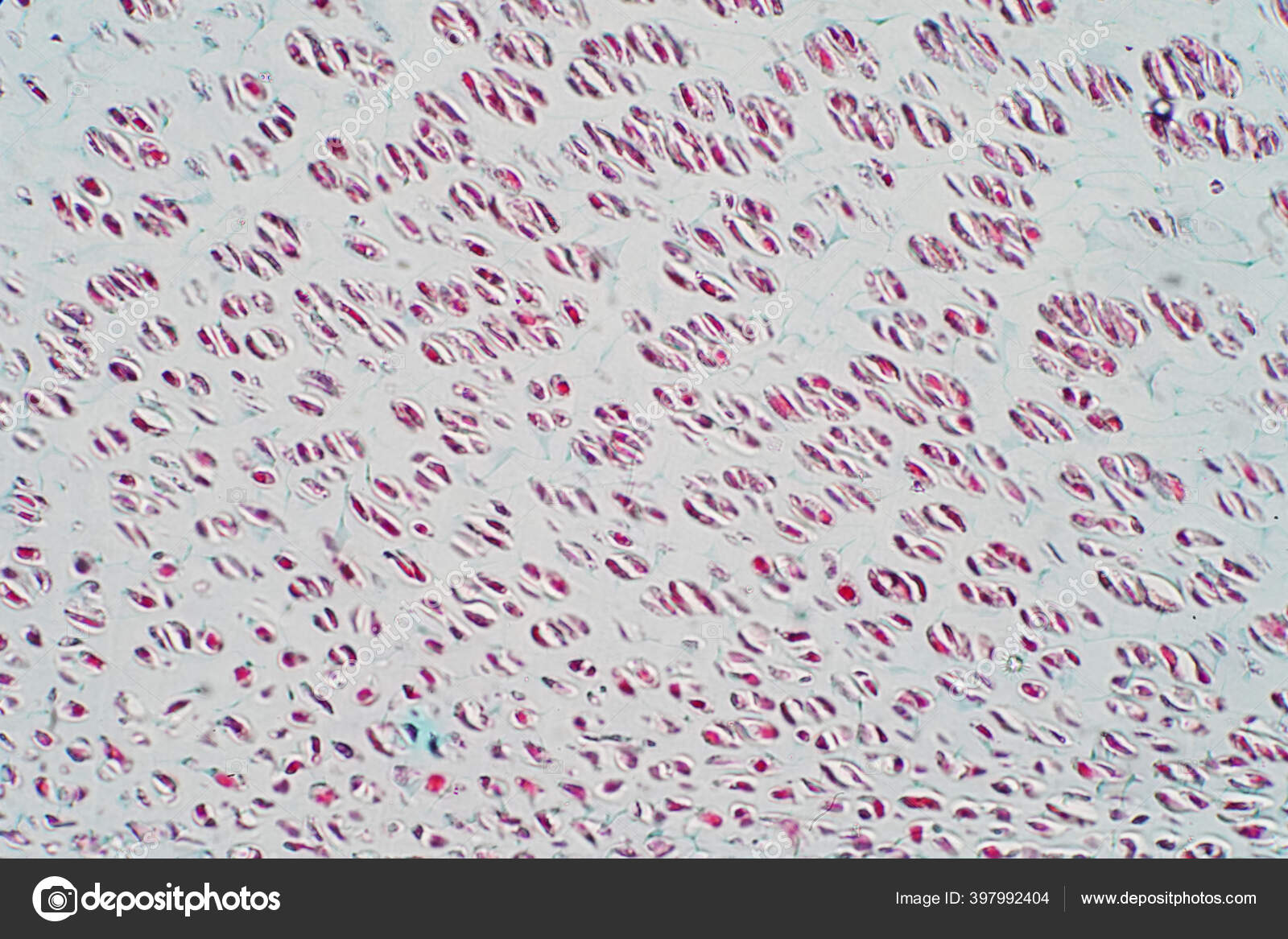
Cross Section Human Cartilage Bone Microscope View Human Histological Physiology Stock Photo Image By C Tonaquatic19 397992404 from st4.depositphotos.com Compact bone cross section courtesy: The finished bone section will be bonded to a microscope slide and so the first step is to grind flat and polish the part of the bone that will be glued to the slide. Cross section performed on focused electon beam (fib) microscope at the university of kentucky's electron microscopy center. Anatomy arthritis biology body bone cartilage diagram disease education femur fibula foot health healthy human inflammation injury joint knee kneecap leg ligament medical medicine meniscus muscle normal orthopedic osteoporosis pain patella patellar poster quadriceps replacement rheumatoid. Add or remove body systems. The concept of a nuclear cross section can be quantified physically in terms of characteristic area where a larger area means a larger probability of interaction. Bones are rigid organs that support and protect various organs of the body, produce red and white blood cells and store minerals. Bone marrow aspiration uses a hollow needle to remove a small sample (about 1 ml) of bone marrow for examination under a microscope.
Bone cross section — stock image & photo.
Jump to navigation jump to search. Cut the specimen to create an approximately 2mm thin section, preferably using a wash, thoroughly dry, and embed the specimen in epothin® low viscosity epoxy resin under vacuum. Bone basics and bone anatomyhave you ever seen fossil remains of dinosaur and ancient human bones in textbooks, television, or in person if you were to look at it in under a microscope, it would look a lot like your kitchen sponge. Bone cross section illustrations & vectors. This simply involves placing a section of the bone on the microscope stage and viewing. Cross section human cartilage bone under microscope view for education histology. They build the entire picture, improve your understanding, consolidate the information and facilitate recall. When the light that enters the condenser is polarized by placing a polarizer in the filter holder and a second, crossed polarizer at the image plane. Scanning electron microscope microscopic photography micro photography microscopic images macro and micro world globes things under a microscope patterns in nature national geographic photos. There are two ways to study bone histology. Microscopic footage of what a human femur bone looks like under the microscope. The infobox for that structure appears on the left of the screen. Cross section of ground compact bone.
This simply involves placing a section of the bone on the microscope stage and viewing. When the light that enters the condenser is polarized by placing a polarizer in the filter holder and a second, crossed polarizer at the image plane. Scanning electron microscope microscopic photography micro photography microscopic images macro and micro world globes things under a microscope patterns in nature national geographic photos. The jeol ion beam cross section polisher (cp) is widely used for preparing pristine samples prior to high resolution imaging and elemental analysis with the scanning electron microscope (sem). This slide showing a cross section of the mammalian trachea (wind pipe) contains examples of several different kinds of tissues.
Bone Cross Section Microscope Woodpeckers Bone Marrow Aspiration Uses A Hollow Needle To Remove A Small Sample About 1 Ml Of Bone Marrow For Examination Under A Microscope from tse4.mm.bing.net Bone basics and bone anatomyhave you ever seen fossil remains of dinosaur and ancient human bones in textbooks, television, or in person if you were to look at it in under a microscope, it would look a lot like your kitchen sponge. Bone cross section — stock image & photo. Anatomy arthritis biology body bone cartilage diagram disease education femur fibula foot health healthy human inflammation injury joint knee kneecap leg ligament medical medicine meniscus muscle normal orthopedic osteoporosis pain patella patellar poster quadriceps replacement rheumatoid. The microscopic cross section represents the effective target area of a single target nucleus for an incident particle. This slide showing a cross section of the mammalian trachea (wind pipe) contains examples of several different kinds of tissues. Both types of bone marrow are enriched with blood vessels and capillaries.2. To download this image, create an account. Cross section human cartilage bone under microscope view for education histology.
As shown in figure 2.
To download this image, create an account. They build the entire picture, improve your understanding, consolidate the information and facilitate recall. To start, select the structure on the model. Cross section performed on focused electon beam (fib) microscope at the university of kentucky's electron microscopy center. Bone basics and bone anatomyhave you ever seen fossil remains of dinosaur and ancient human bones in textbooks, television, or in person if you were to look at it in under a microscope, it would look a lot like your kitchen sponge. Pores are filled with marrow, nerves, and blood vessels that carry. A uniform cross section is the cross section of the solid, parallel to base, such that the resulting figure has the same shape and size as that of the base of the figure.more about uniform cross sectionsolids like pyramids and cones have slant heights and hence do not have uniform cross. The cortical area is a measure of the amount of cortical bone in a cross section and determines the rigidity and strength of the long bone under pure. Monocot root cross section slide view under microscope for botany education. The concept of a nuclear cross section can be quantified physically in terms of characteristic area where a larger area means a larger probability of interaction. The microscopic cross section represents the effective target area of a single target nucleus for an incident particle. Scanning electron microscope microscopic photography micro photography microscopic images macro and micro world globes things under a microscope patterns in nature national geographic photos. A cross section of a compact bone shows concentric circles called lamellae.
Newly designed microscope slide for cutting and viewing a quick cross section. Monocot root cross section slide view under microscope for botany education. Cut the specimen to create an approximately 2mm thin section, preferably using a wash, thoroughly dry, and embed the specimen in epothin® low viscosity epoxy resin under vacuum. The units are given in barns or cm2. To download this image, create an account.
Cross Section Human Cartilage Bone Under Stock Photo Edit Now 1072783703 from image.shutterstock.com Cross section performed on focused electon beam (fib) microscope at the university of kentucky's electron microscopy center. Pores are filled with marrow, nerves, and blood vessels that carry. Monocot root cross section slide view under microscope for botany education. Both types of bone marrow are enriched with blood vessels and capillaries.2. The units are given in barns or cm2. Note that the bone matrix is deposited in concentric layers called lamellae. From wikimedia commons, the free media repository. When the light that enters the condenser is polarized by placing a polarizer in the filter holder and a second, crossed polarizer at the image plane.
As shown in figure 2.
Add or remove body systems. Where speed is essential, such as in surgical biopsies for cancer. Bone cross section — stock image & photo. Bone basics and bone anatomyhave you ever seen fossil remains of dinosaur and ancient human bones in textbooks, television, or in person if you were to look at it in under a microscope, it would look a lot like your kitchen sponge. They build the entire picture, improve your understanding, consolidate the information and facilitate recall. The finished bone section will be bonded to a microscope slide and so the first step is to grind flat and polish the part of the bone that will be glued to the slide. Cross section of ground compact bone. Bones are rigid organs that support and protect various organs of the body, produce red and white blood cells and store minerals. The microscopic cross section represents the effective target area of a single target nucleus for an incident particle. Department of histology, jagiellonian university medical under the stereo microscope (and depending on the section of the bone under. When the light that enters the condenser is polarized by placing a polarizer in the filter holder and a second, crossed polarizer at the image plane. To start, select the structure on the model. Cross section human skin tissue under microscope view.
Peripheral nuclei, connective tissue and myofibers bone cross section. Bone cross section illustrations & vectors.